Quick answer
Cell therapy is used for ankle cartilage damage (osteochondral lesions of the talus, or OLTs) mainly as an add-on to surgery, most often with bone marrow aspirate concentrate (BMAC), not as a proven stand-alone cure. In one 101-patient study, adding BMAC to microfracture cut the revision rate from 28.8% to 12.2%, but the groups were not randomized and the authors called for larger trials. Standard treatment still comes first: protected weight-bearing and physiotherapy, then surgery such as microfracture, which gives good or excellent results in over 70% of patients.
Key facts at a glance
- What it is: damage to the cartilage and the bone under it on the top of the ankle bone (talus), most often after a sprain.
- First-line care: rest, restricted weight-bearing and physiotherapy; surgery if pain persists.
- Cell therapy role: mostly BMAC or cultured cells placed during surgery, to help the repair tissue heal.
- Evidence: promising but limited; no large randomized trial proves a clear benefit yet.
- Best candidates: usually people with small to medium lesions who have not improved with non-surgical care.
What is an osteochondral lesion of the talus (ankle cartilage damage)?
An osteochondral lesion of the talus (OLT) is an injury to the cartilage and the bone beneath it on the top of the ankle bone (the talus), which can include softened cartilage, a cyst in the bone or a fractured fragment (FootCareMD). Cartilage has very little blood supply, so it heals poorly once damaged. Doctors also call it a talar dome lesion or osteochondritis dissecans of the talus.
What causes ankle cartilage damage?
Up to 85% of OLTs follow an ankle injury, usually a sprain in which the talus strikes another bone of the ankle. Some people have no injury history, and inner-side lesions can sometimes develop during growth (FootCareMD).
What are the symptoms?
- Ankle pain that lasts for months after a sprain despite rest and elevation.
- Swelling, a catching or locking feeling, and a sense of instability.
- Pain on the outer or inner side of the ankle, often worse with activity.
Severe locking may mean a large lesion or a loose fragment in the joint.
How is it diagnosed?
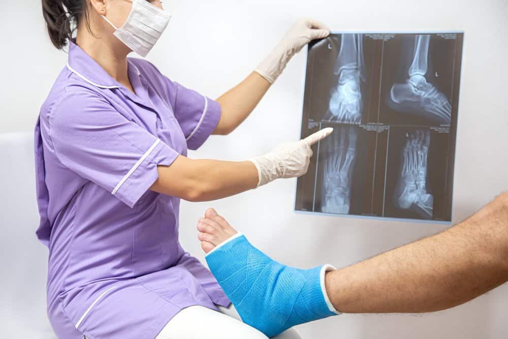
A foot and ankle specialist suspects an OLT from your history and examination. Standard X-rays show some lesions, but CT or MRI is often needed to confirm it and to measure its size and depth (FootCareMD). Lesion size and location, any cyst, and whether the ankle is unstable all affect the treatment choice.
If an old sprain still hurts, read Viezec’s guides on stem cell therapy for cartilage repair, stem cell treatment for cartilage repair in India and stem cell therapy for tendon and ligament injuries.
What are the conventional treatments for ankle cartilage damage?
Conventional treatment starts with protecting the ankle and moves to surgery only if pain continues. Which step you need depends on whether the lesion is stable, how large it is and whether the ankle is unstable.
| Treatment | What it involves | When it is used | Main limits |
|---|---|---|---|
| Protection and rehabilitation | Cast or boot, restricted weight-bearing, gradual return to walking, then physiotherapy; anti-inflammatory tablets and a brace if the ankle is unstable (FootCareMD, Foot Health Facts) | Stable lesions, no loose fragment; first step for most patients | Full pain relief in fewer than half of cases (FootCareMD) |
| Debridement and fixation | Arthroscopic removal of damaged tissue, or pinning or screwing a loose fragment back | Loose or unstable fragments | Does not rebuild lost cartilage |
| Microfracture or drilling | Small holes in the bone so blood and marrow cells form repair tissue | Small to medium lesions that did not respond to rest | Repair tissue is not normal cartilage; larger lesions do worse. Over 70% of patients have good or excellent results after debridement and microfracture (FootCareMD) |
| Bone grafting of a cyst | Filling a bone cyst under the cartilage | Lesions with a cyst | Needs a longer protected recovery |
| Bone and cartilage transfer or grafting | Moving healthy bone and cartilage into the defect | Larger lesions or when microfracture failed | Longer recovery; graft failure and donor-site problems are possible |
| Correcting related problems | Treating ankle instability, impingement, a high-arched foot or tight calf muscles during surgery | When these contribute to the lesion | Adds to surgery and rehab time |
Smaller lesions generally do best, and most procedures need several weeks to months of restricted weight-bearing, with longer recovery for grafting and transfer (FootCareMD).
What conventional treatment cannot do
None of these options reliably regrows normal hyaline cartilage, and a damaged ankle can go on to arthritis. That is why surgeons study biological add-ons such as bone marrow-derived cells.
Viezec’s pages on stem cell therapy vs surgery and importance of physical therapy following stem cell procedures explain how cell therapy fits alongside surgery and rehab.
How is cell therapy used for ankle cartilage damage?
Cell therapy for ankle cartilage lesions is used mainly during or alongside a repair procedure, to supply cells and growth signals that help the new tissue form. The goal is to improve the quality of the repair tissue, not to inject cells into a healthy ankle and expect cartilage to regrow on its own.
What types of cell-based treatment are used?
- Bone marrow aspirate concentrate (BMAC): marrow is drawn from the patient’s own bone, spun to concentrate the cells and growth factors, and applied to the lesion, often together with microfracture and a fibrin glue carrier. This is the most studied option for talar lesions (cohort study). See Viezec’s pages on bone marrow aspirate injections for joint osteoarthritis and autologous bone marrow-derived cell therapy.
- Scaffold-assisted repair (such as AMIC and collagen scaffolds): microfracture combined with a collagen membrane or scaffold that holds the clot and marrow cells in place. Studies report good pain and function scores, and a collagen-scaffold study found similar results to microfracture at 1 year with higher patient satisfaction (Singapore General Hospital).
- Autologous chondrocyte implantation (ACI): the patient’s own cartilage cells are grown in the laboratory and implanted in a second operation. It is effective at stimulating hyaline-like cartilage but is staged and costly (Singapore General Hospital).
- Platelet-rich plasma (PRP) and mesenchymal stem cell injections: used as supportive injections for pain and healing, with less evidence specific to the talus. Compare them in stem cell therapy vs PRP therapy and read about bone marrow-derived mesenchymal stem cells.
How might cell therapy help?
Marrow cells and platelets release growth factors that support healing and may improve how the microfracture clot matures into repair tissue. This is a proposed mechanism, and it has not been shown to turn the repair tissue into normal hyaline cartilage in every patient.
For other foot and ankle uses, see Viezec’s potential of stem cell therapy for feet and stem cell treatment for sports injuries.
What does the research say about cell therapy for ankle cartilage lesions?
The research shows encouraging results when cells or scaffolds are added to surgery, but the best studies are small, non-randomized or short-term. Four findings matter most.
- BMAC with microfracture: In a prospective study of 101 patients followed for at least 36 months, both microfracture alone (52 patients) and microfracture with BMAC (49 patients) improved pain, quality of life and sport participation. Revision surgery was needed in 28.8% of the microfracture group and 12.2% of the BMAC group. The groups were treated in different years rather than randomized (level III evidence), and the authors asked for larger randomized trials (cohort study).
- Overall BMAC evidence: A 2023 systematic review and meta-analysis of BMAC added to bone marrow stimulation for talar lesions is titled “Limited evidence in support of bone marrow aspirate concentrate as an additive” (PubMed). Treat BMAC as promising but not proven.
- Microfracture alone has limits: A Singapore review reported excellent or good results in 83% to 89% of patients at up to 2 years, yet one study found 40% of patients had abnormal cartilage healing on second-look arthroscopy, because the repair tissue is fibrocartilage rather than hyaline cartilage (Singapore General Hospital).
- Scaffold-assisted repair: Reported AMIC follow-up of 2 to 8 years showed pain falling from 6.4 to 1.4 out of 10, 79% returning to sport and defect filling on MRI in over 88% (Singapore General Hospital). A scaffold study using the same source reported similar 1-year scores to microfracture with higher satisfaction.
Does lesion size change the evidence?
Yes. Bone marrow stimulation works best for smaller lesions, with the Singapore study including only lesions under 1.5 cm², and larger or cystic lesions often need grafting such as osteochondral autograft transfer. In one 20-patient series with about 12-year follow-up, graft transfer raised the AOFAS ankle score from 60.4 to 86.2, but 14% had lasting knee donor-site pain (Journal of Joint Diseases and Related Surgery).
Is stem cell therapy approved for ankle cartilage damage?
Stem cell and BMAC use for ankle cartilage is generally still investigational or off-label, and the US FDA states that no regenerative medicine therapy is approved for orthopedic conditions such as osteoarthritis (FDA). Ask any clinic how its treatment is regulated.
For Viezec’s own discussion of outcomes, see stem cell success rate and how long stem cell injections last for knee pain.
Cell therapy vs conventional treatment for ankle cartilage lesions: comparison
Cell therapy is an add-on to proven repair techniques, not a replacement for them. This table shows how each option fits.
| Option | Best suited to | What the evidence shows | Main limits |
|---|---|---|---|
| Protection and physiotherapy | Stable lesions, first step for most | Full pain relief in fewer than half of cases | Many patients still need surgery |
| Microfracture alone | Small lesions that failed non-surgical care | Good or excellent results in over 70% (83% to 89% in a Singapore review) | Fibrocartilage repair; larger lesions do worse |
| Microfracture plus BMAC | Small to medium lesions where a biological boost is wanted | Revision rate 12.2% vs 28.8% in a 101-patient non-randomized study; wider evidence rated limited | Not randomized; marrow collection adds a procedure; investigational in many countries |
| Scaffold-assisted repair (AMIC, collagen) | Lesions needing a framework for the clot | Good scores and MRI defect filling in case series | Often needs open surgery; longer-term data needed |
| Autologous chondrocyte implantation | Larger or failed lesions | Stimulates hyaline-like cartilage | Two operations and high cost |
| Bone and cartilage transfer (OAT) | Large, uncontained or cystic lesions | Good to excellent in about 90% at around 12 years in one 20-patient series | Often needs an ankle bone cut; knee donor-site pain in 14% |
| Injections alone (PRP or stem cells) | Mild pain, or when surgery is not wanted | Little ankle-specific research | Not shown to repair a defined lesion |
Sources: FootCareMD, cohort study, Singapore General Hospital, Journal of Joint Diseases and Related Surgery.
To see how Viezec compares non-surgical and surgical routes, read stem cell therapy vs surgery and prp and stem cell therapy for partial knee replacement.
Is cell therapy for ankle cartilage damage safe?
When cells are taken from your own body and used during a supervised surgery, the added risk is generally small, but no procedure is risk-free. Most risks come from the surgery and from poorly regulated products, not from your own marrow cells.
What are the possible risks?
- Surgical risks: anesthesia complications, infection, nerve or blood vessel damage, bleeding and blood clots. Infection is uncommon after arthroscopic surgery, but open or transfer procedures carry added risk, including graft failure (FootCareMD).
- Marrow collection site: soreness, bruising or bleeding where the marrow is taken.
- Persistent pain or a repair that fails, which may need further treatment.
- Unregulated products: the FDA warns that unapproved stem cell, Wharton’s jelly and exosome products have been linked to infections, tumour formation and neurological events, and that offers made outside a regulated trial may be illegal (FDA).
Who may be a candidate, and what does recovery look like?
The people most likely to be considered for cell-assisted repair are those with an MRI-confirmed lesion, ongoing pain after months of non-surgical care, and a surgeon who judges that a repair is needed. The decision belongs to a foot and ankle specialist, because the lesion size, cysts and ankle stability decide the technique.
How to lower the risk
- Have an MRI and a foot and ankle specialist assess the lesion before choosing any treatment.
- Treat related problems such as ankle instability or a high-arched foot, or the repair may fail.
- Prefer your own cells (autologous) processed under documented quality control.
- Ask for a written risk list, and avoid any clinic that guarantees cartilage regrowth.
Read Viezec’s pages on safety and adverse effects of stem cell treatment, stem cell therapy safety and quality control tests.
Who may be a candidate, and what does recovery look like?
The people most likely to be considered for cell-assisted repair are those with an MRI-confirmed lesion, ongoing pain after months of non-surgical care, and a surgeon who judges that a repair is needed. The decision belongs to a foot and ankle specialist, because the lesion size, cysts and ankle stability decide the technique.
Who may be considered
- Adults with persistent ankle pain, swelling or catching after a sprain, confirmed on MRI or CT.
- Patients whose stable lesion did not improve with a cast or boot, physiotherapy and medicines.
- Active people who want to protect the joint and return to sport.
Who should be cautious or look at other options first
- People with advanced ankle arthritis, where cartilage repair is less likely to help.
- Those with untreated ankle instability, impingement or a high-arched foot, which may need correction first.
- Patients with active infection, uncontrolled diabetes, smoking or poor circulation, which can slow healing.
- Anyone with a large or cystic lesion, where grafting may be the better evidence-backed choice.
What does recovery involve?
- Protection: most treatments need immobilization and restricted weight-bearing for several weeks to months, longer after grafting or transfer (FootCareMD).
- Gradual loading: a staged return to walking, guided by your surgeon.
- Physiotherapy: range of motion, calf and ankle strengthening, then balance and sport-specific work. See the importance of physical therapy following stem cell procedures.
- Follow-up imaging if pain returns or the ankle swells.
For athletes, Viezec’s guides on how athletes benefit from stem cell therapy and stem cell therapy restoring mobility to athletes add context.
How to evaluate a clinic offering cell therapy for the ankle
- Ask whether the cells are your own, how they are collected, processed and tested, and whether they are used with surgery or injected alone.
- Ask for your lesion size and what the evidence is for that size.
- Ask who will do the procedure and the rehabilitation, and for the written risk list.
- Be cautious if a clinic promises cartilage regrowth or a cure.
To discuss your MRI and reports, contact the Viezec team. International patients can start with the international patient flow chart.
Frequently asked questions
Some lesions improve with a cast or boot, restricted weight-bearing and physiotherapy, but full pain relief occurs in fewer than half of cases. Cartilage has a poor blood supply, so persistent pain after several months usually needs a specialist review.
It is damage to the cartilage and underlying bone on top of the ankle bone (the talus), most often after an ankle sprain. It can cause long-lasting pain, swelling, catching and instability.
Cell therapy, mainly bone marrow aspirate concentrate used with surgery, may improve how the repair tissue heals. It has not been proven to regrow normal cartilage, and the evidence is limited to small or non-randomized studies.
One 101-patient study found fewer revision surgeries with BMAC (12.2% vs 28.8%), but the groups were not randomized. A 2023 meta-analysis of the question is titled “limited evidence”, so BMAC is promising but unproven.
Not always. Stable lesions often start with a cast or boot and physiotherapy. Surgery is considered if symptoms continue, if a fragment is loose, or if the lesion is large.
Most treatments need several weeks to months of restricted weight-bearing, and grafting or transfer procedures take longer. Your surgeon will set the timeline for your lesion.
Injections of PRP or stem cells are offered for ankle pain, but there is little ankle-specific research showing they repair a defined lesion. Treat them as supportive, and confirm the diagnosis with an MRI first.
The US FDA states that no regenerative medicine therapy is approved for orthopedic conditions such as osteoarthritis, and unapproved products offered outside a trial can be unsafe. Ask any clinic how its treatment is regulated.
Ask whether the cells are your own, how they are processed and tested, whether they are used with surgery, what your lesion size means for results, and what the written risks are. Avoid guarantees of regrowth.
Conditions We Treat
Explore stem cell treatment options in India